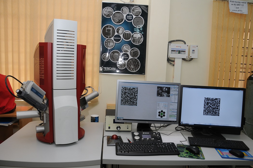
Field Emission Scanning Electron Microscope
Technical and Characterization Specifications:
- 2X to10,00,000X Magnification
- High vacuum and low vacuum ( up to 500 Pa) imaging
- IR camera for chamber view
- Everhart-Thornley type detectors with YAG scintillators
- Chamber SE detector, Resolution-1.2 nm at 30 kV (SE: Secondary electrons)
- In-Beam SE detector,Resolution-1.0 nm at 30 kV
- In-Beam backscattered SE detector , Resolution-2.0 nm at 15 kV
- Low vacuum SE detector with differentially pumped detection chamber and a dedicated turbo molecular pump , Resolution-3.0 nm at 30 kV
- Beam deceleration mode (BDM) with in-Beam annular SE detector for thin films, semiconductors and also for specimens prone to radiation damages, Resolution-1.8 nm at 1kV
- Air cooled column
- Pneumatic anti-vibration suspension system
- Accelerating voltage- 50 V to 30 kV in steps of 10 V
- Probe current 2 pA to 200 nA
- Probe current detector
- Field of view- 6.4 mm at WD 10 mm and 20 mm at WD 30 mm
- 20 ns to 10 ms per pixel scanning speed (Adjustable continuously)
- Selectable image frame size up to 8192 X 8192 pixels
- Point & line scan, image rotation & shift and tilt compensation
- Sample stage movements :
- Motorized X,Y,Z = 80 mm, 60 mm, 47 mm respectively
- Rotation = 360 deg –motorized
- Tilt = -80o to +80o -motorized
- Compucentric stage
- Multiple specimen holder
- Fixed Scanning transmission electron microscope (STEM) Detector –
- Sample prepared using standard TEM grids for sample insertion
- Resolution in high vacuum- 0.8 nm at 30 kV
- Bright field and dark field imaging